People
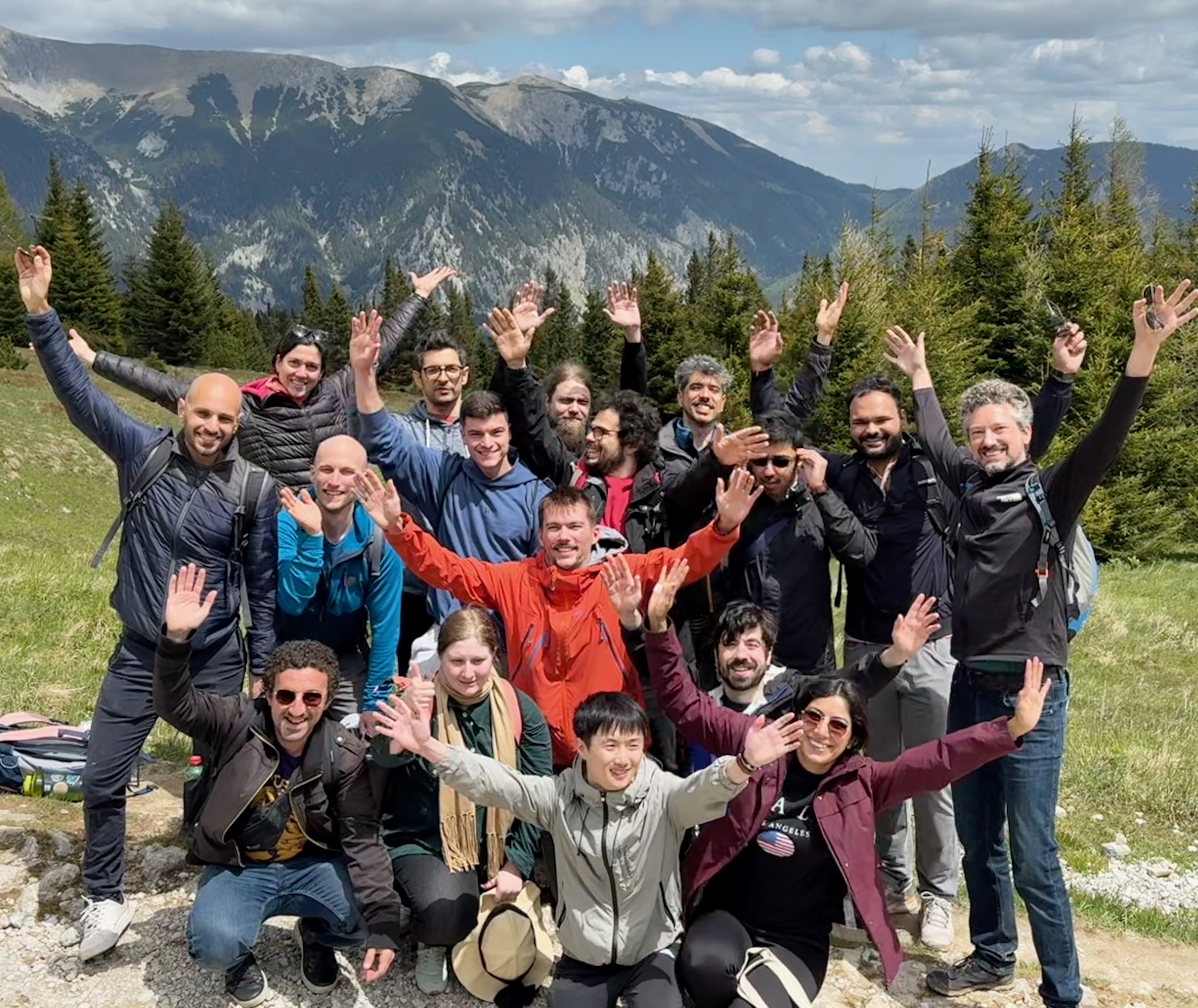
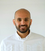
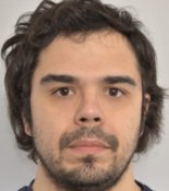
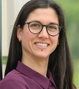
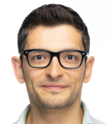
Group Members in Vienna


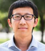
| Jonas Ries | |
|---|---|
| Since 2023 | Professor for Advanced Microscopy and Cellular Dynamics at the Max Perutz Labs, University of Vienna. |
| 2012 - 2023 | Group leader at the EMBL in Heidelberg |
| 2009 - 2012 | Postdoc at ETH Zürich with Vahid Sandoghdar and Helge Ewers: Novel labeling schemes for superresolution microscopy |
| 2005 - 2008 | PhD at TU Dresden with Petra Schwille: Advanced fluorescence correlation methods to study membrane dynamics |



Open positions
With our move to the Max Perutz Labs at the University of Vienna we have competitive positions for applicants with a background in physics, programming or biology on all levels. Please contact Jonas directly.
Have a look at our job advertisements at the Max Perutz Labs.
If you want to join as a PhD student, please note that admission is exclusively via the Vienna BioCenter PhD program.
Alumni
| Name | Position | Topic | Period | Current affiliation |
|---|---|---|---|---|
| Takahiro Deguchi | Postdoc | MINFLUX | 06.2020 - 12.2024 | EMBL Heidelberg, Zimmermann group |
| Soheil Mojiri | Postdoc | Cryo-SMLM | 01.2022 - 12.2024 | EMBL Heidelberg, Mahamid group |
| Hao Sha | Visiting PhD student | Deep learning | 10.2024 - 8.2025 | |
| Shantanu Yadav | Intern | Deep learning | 5.2025 - 7.2025 | |
| Michaela Janka | Master student | Endocytosis | 6.2024 - 7.2025 | |
| Sidak Singh Grewal | Intern | Microscopy | 5.2025 - 7.2025 | |
| Pau Garcia | Master student | Kinesin tracking | 11.2024 - 6.2025 | |
| Phylicia Kidd | Visiting Scientist | Expansion microscopy | 01.2024 - 11.2024 | IMP Vienna |
| Lydia Steiner | Intern | MINFLUX | 08.2024 - 11.2024 | |
| Lucas-Raphael Müller | PhD student | Deep learning for SMLM | 05.2024 - 07.2024 | |
| Gabin Agbale | Master student | Deep learning for SMLM | 04.2024 - 09.2024 | |
| Lukas Scheiderer | Postdoc | MINFLUX | 02.2024 - 07.2024 | |
| Valerie Segatz | Intern | MINFLUX | 04.2024 - 07.2024 | |
| Alejandro Linares | PhD student | Endocytosis | 09.2023 - 06.2024 | TU Wien |
| Andrea Todorovic | Intern | Kinesin tracking with MINFLUX | 01.2024 - 03.2024 | |
| Arthur Jaques | Master student | Deep learning for SMLM | 05.2023 - 03.2024 | |
| Christopher Heidebrecht | Master student | MINFLUX tracking | 03.2023 - 03.2024 | MPI for multidisciplinary sciences |
| Binnur Oezay | Intern | MINFLUX tracking | 11.2023 - 02.2024 | |
| Ajit Roy | Master student | Construction of a microscope | 04.2023 - 01.2024 | |
| Anna Gergely | Intern | 06.2023 - 10.2023 | University of Heidelberg | |
| Sara Klingelhoefer | Intern | SMLM on endocytosis | 02.2023 - 06.2023 | University of Heidelberg |
| Aline Tschanz | PhD student | SMLM on endocytosis | 10.2018 - 10.2023 | |
| Yu-Le Wu | PhD student, bridging postdoc | SMLM data analysis methods | 10.2017 - 04.2023 | DKFZ |
| Timon Andre | Postdoc | SMLM on endocytosis | 10.2022 - 05.2023 | EMBL Heidelberg |
| Marc Molina Jordan | Visiting Scientist | Nuclear Pore Complex | 02.2023 - 05.2023 | IBEC Barcelona |
| Philipp Hoess | PhD student, laboratory officer | SMLM on yeast endocytosis | 09.2016 - 01.2023 | Sanofi |
| Franziska Fichtner | Intern | MINFLUX tracking | 10.2022 - 12.2022 | University of Heidelberg |
| Arnas Zubas | Intern | SMLM of mammalian CME | 07.2022 - 11.2022 | University of Heidelberg |
| Michal Skruzny | Research Scientist | Dual-color SMLM of endocytosis in yeast | 12.2021 - 10.2022 | Zeiss Microscopy |
| Lukas Voos | Intern | Dual-color SMLM of endocytosis in yeast | 04.2022 - 10.2022 | University of Heidelberg |
| Saeed Azizi | Intern | Primed conversion in yeast | 12.2021 - 10.2022 | Heidelberg Instruments |
| Sheng Liu | PostDoc | 4Pi SMLMS & PSF learning | 06.2020 - 06.2022 | University of New Mexico |
| Angelica Maria Estrada Pacheco | Research Technician | SMLM of T cells & DNA origami | 01.2020 - 06.2022 | |
| Eva-Maria Schentarra | Intern | MINFLUX tracking | 10.2021 - 04.2022 | University of Heidelberg |
| Tomas Noordzij | Intern & Visiting Scientist | Endocytosis in Drosophila | 01.2020 - 10.2020 & 03.2021 - 03.2022 | University of Utrecht |
| Lisa Schmidt | Intern | MINFLUX tracking | 05.2021 - 09.2021 | |
| Jonas Hellgoth | Intern | Multi-color SMLM & PSF learning | 04.2021 - 08.2021 | |
| Ulf Matti | Research Technician | The NPC as imaging standard, multi-color SMLM, supporting the lab in all possible ways,... | 07.2012 - 06.2021 | Abberior Instruments, Göttingen |
| Jervis Thevathasan | PhD Student & PostDoc | SMLM of α-synuclein, the NPC as imaging standard | 09.2015 - 06.2021 | |
| Veronika Chevyreva | Visiting PhD Student | Optimization of dual-color SMLM in yeast | 12.2020 - 04.2021 | IFOM, Milan, Italy |
| Jan-Niklas Wittemann | Intern | Dual-color SMLM of mammalian CME | 12.2020 - 04.2021 | |
| Marija Minzburg | Intern | Modeling of 4 Pi PSF | 11.2020 - 02.2021 | |
| Lisa Nechyporenko | Intern | Analysis of kinetochore super-resolution data & exploring fluorescent proteins in yeast | 03.2020 - 08.2020 | University of Heidelberg |
| Vincent Casamayou | Intern | VR visualization of super-resolution data | 03.2020 - 08.2020 | |
| Joran Deschamps | PhD Student & Scientific Officer | SALM, automation of SMLM, software development (EMU, FPGA, and device adapters for Micro-Manager),... | 09.2013 - 07.2020 | Human Technopole, Milan, Italy |
| Leonard Krupnik | Master Student | SMLM of α-synuclein | 10.2019 - 05.2020 | Empa, St. Gallen, Switzerland |
| Robin Diekmann | PostDoc | Ratiometric SMLM, camera calibration, slowSTORM, 4 Pi microscopy | 01.2018 - 04.2020 | LaVision BioTec, Bielefeld |
| Andreas Schoenit | Intern | The NPC as imaging standard | 10.2019 - 03.2020 | MPI Medical Research, Heidelberg |
| Maurice Kahnwald | Master Student | The NPC as imaging standard | 09.2018 - 11.2019 | FMI, Basel, Switzerland |
| Anindita Dasgupta | Master Student | Supercritical angle localization microscopy | 09.2018 - 11.2019 | Institute of Applied Optics and Biophysics, Jena |
| Yiming Li | PostDoc | 4 Pi microscopy & experimental PSF modeling | 01.2016 - 10.2019 | Southern University of Science and Technology, Shenzhen, China |
| Amir Rahmani | Intern | Optimizing Dual-Color SMLM | 07.2019 - 09.2019 | Shahid Beheshti Universiti, Tehran, Iran |
| Cheng-Yu Huang | Intern | PSF Modeling | 06.2019 - 09.2019 | Cambridge University, UK |
| Eric Maurer | Intern | Super-resolution microscopy using small tags | 06.2019 - 08.2019 | University of Heidelberg |
| Konstanty Cieśliński | PhD Student & PostDoc | Super-resolution imaging of the yeast kinetochore | 10.2013 - 12.2018 | DKFZ, Heidelberg |
| Alejandro Colchero | Master Student | Optimizing dual-color SMLM | 04.2018 - 09.2018 | University of Madrid, Spain |
| Sudheer Kumar Peneti | Master Student | Quantifying labeling efficiency in SMLM | 04.2018 - 07.2018 | |
| Raika Karimi | Intern | 4 Pi software development | 04.2018 - 06.2018 | Montreal, Canada |
| Julia Botta | Intern | Counting by SMLM and kinetochore | 02.2018 - 04.2018 | University of Heidelberg |
| Daniel Heid | Intern | Quantifying labeling efficiency in SMLM | 01.2018 - 04.2018 | EMBL Heidelberg |
| Li-Ling Yang | PostDoc | Inverted lattice light-sheet microscope | 06.2014 - 12.2017 | Charité Berlin |
| Markus Mund | PhD Student & PostDoc | Endocytosis | 09.2012 - 12.2017 | University of Geneva, Switzerland |
| Sarah Hörner | Master Student | Counting with SMLM | 06.2017 - 12.2017 | Heidelberg University |
| Elena Buglakova | Intern | Modeling of 4Pi-PSF | 07.2017 - 09.2017 | EMBL Heidelberg |
| Daniel Schroeder | Master Student | Microscopy development | 04.2017 - 08.2017 | FU Berlin |
| Krishna Kasuba | Intern | Protein counting by SMLM | 10.2016 - 05.2017 | ETH Zürich, Switzerland |
| Johanna Mehl | Intern | Superresolution imaging of endocytosis | 10.2016 - 03.2017 | ETH Zürich, Switzerland |
| Jan van der Beek | Intern | Superresolution imaging of endocytosis | 12.2014 - 12.2016 | Utrecht University, Netherlands |
| Jooske Monster | Intern | Superresolution imaging of endocytosis | 09.2016 - 12.2016 | Utrecht University, Netherlands |
| Joanna Zareba | Intern | Fluorophores | 06.2016 - 10.2016 | University of Zürich, Switzerland |
| Katharina Lindner | Intern | Kinetochore | 02.2016 - 05.2016 | Heidelberg University |
| Silvia Seidlitz | Bachelor Student | Lattice light sheet microscopy | 11.2015 - 04.2016 | DKFZ, Heidelberg |
| Manuel Reitberger | Master Student | SMLM counting | 09.2015 - 04.2016 | DKFZ, Heidelberg |
| Ronny Sczech | Software Developer | SMLM software development | 04.2015 - 02.2016 | Leipzig |
| Tooba Quidwai | Intern | Superresolution imaging of platelets | 04.2014 - 08.2015 | Edinburgh, UK |
| Abhijit Marar | Master Student | Fitting software | 11.2014 - 07.2015 | Georgia Institute of Technology |
| Andreas Rowald | Intern | Microscopy development | 09.2014 - 02.2015 | EPFL, Switzerland |
| Rohit Prakash | Intern | Superresolution imaging of endocytosis | 11.2013 - 01.2015 | Rochester University, US |
| Nagarajan Chandramohan | Intern | Programming | 01.2014 - 11.2014 | PEPperPRINT GmbH |
| Sven Spachmann | Bachelor student | Co-localization in superresolution | 05.2014 - 08.2014 | Heidelberg University |
| Sunil Kumar Dogga | Intern | Endocytosis | 11.2013 - 03.2014 | Sanger Institute, Cambridge, UK |
| Sanchari Datta | Intern | Endocytosis | 06.2013 - 07.2013 | |
| Meet Mukesh Paswan | Intern | Programming | 06.2013 - 07.2013 | |
| Johannes Bues | Intern | Superresolution in yeast | 04.2013 - 05.2013 |